

No, you cannot definitively identify toxic algae by visual inspection alone. While certain characteristics like a “spilled paint” appearance or “pea soup” color are strong indicators of a cyanobacterial bloom, non-toxic green algae can exhibit similar phenotypes. Conversely, lethal concentrations of cyanotoxins can persist in water that appears clear to the naked eye. Laboratory analysis, such as ELISA or LC-MS/MS testing, remains the only scientifically valid method for determining toxicity.
Your eyes can lie, but the science doesn’t. Learn the visual red flags of toxic blooms. You can’t always see toxins, but you can see the signs of a bloom. Protect your family and pets with our identification guide.
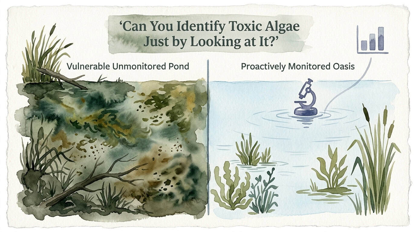
Understanding the limitations of human perception in aquatic environments is critical for risk mitigation. Cyanobacteria, often colloquially termed “blue-green algae,” are complex prokaryotic organisms capable of synthesizing potent secondary metabolites known as cyanotoxins. These toxins represent a significant pharmacological threat to hepatic, neurological, and dermatological systems in mammals. Relying on visual heuristics is a suboptimal strategy for safety; instead, a multi-tiered approach involving field screening and laboratory validation is required to manage a Vulnerable Unmonitored Pond effectively and transition it toward becoming a Proactively Monitored Oasis.
Can You Identify Toxic Algae Just by Looking at It?
Cyanobacterial Harmful Algal Blooms (CyanoHABs) are proliferations of photosynthetic bacteria that outcompete eukaryotic algae under specific environmental conditions, such as high phosphorus loading and thermal stratification. Although they perform photosynthesis like plants, cyanobacteria are structurally distinct. They lack membrane-bound organelles and possess specialized pigments, primarily phycocyanin, which allows them to harvest light at wavelengths that other aquatic plants cannot utilize.

The presence of a bloom does not inherently guarantee the presence of toxins. Research indicates that the genes responsible for toxin production (such as the mcy gene cluster for microcystins) may be present in a population but not actively expressed, or the population may consist of a mix of toxic and non-toxic strains. This biological variability makes visual identification impossible for determining actual health risks. A pond might appear covered in a dense green scum but contain zero detectable toxins, while a nearby lake with high water clarity might harbor dangerous levels of anatoxin-a produced by benthic mats at the bottom.
In real-world scenarios, water resource managers utilize visual cues only as a trigger for further diagnostic testing. These cues serve as an initial “triage” step in a broader monitoring protocol. Because cyanobacteria can regulate their buoyancy using internal gas vesicles, they often aggregate at the surface during calm conditions, creating the high-visibility “blooms” that the public associates with toxicity.
Mechanisms of Visual Deception and Morphological Analysis
Visual assessment of water bodies is frequently confounded by “look-alikes” that possess no toxic potential. Distinguishing between these requires an understanding of the mechanical and biological properties of the organisms involved.
One of the most effective field-level mechanical tests is the “stick test.” Filamentous green algae, such as Spirogyra or Cladophora, possess a cellular structure that allows them to form long, cohesive strands or mats. When a stick is inserted into these mats and lifted, the algae will hang in long, hair-like strings. In contrast, cyanobacteria like Microcystis do not form these structural filaments. When subjected to a stick test, cyanobacteria typically behave like liquid paint or small individual flecks, failing to cling to the stick in a cohesive manner.
Another diagnostic tool is the “jar test.” This utilizes the buoyancy regulation of cyanobacteria. A water sample is collected in a clear glass jar and left undisturbed for 15 to 30 minutes. Because many bloom-forming cyanobacteria (such as Dolichospermum) contain gas vesicles, they will migrate to the surface to form a concentrated layer. True green algae, which are heavier than water and lack buoyancy control, will settle at the bottom of the jar.
Despite these tests, several common phenomena mimic CyanoHABs:

- Pollen: Pine or oak pollen often forms yellow or light-green drifts on water surfaces. Unlike cyanobacteria, pollen is typically granular and concentrated in late spring.
- Duckweed (Lemnoideae): These are tiny flowering plants, not algae. They possess a distinct leaf-like structure (frond) and tiny roots, which are visible upon close inspection.
- Filamentous Green Algae: While unsightly, these often form “clouds” or “mats” that feel metallic or slimy but do not release toxins into the water column.
Analytical Methodologies for Definitive Identification
To move beyond visual speculation, practitioners employ several laboratory techniques, each with specific detection limits and technical requirements.
Microscopy and Taxonomic Enumeration
The primary step in a professional laboratory is microscopic identification. Technicians look for specific morphological traits:
- Microcystis: Forms large colonies of small, spherical cells held together by a mucilaginous matrix.
- Dolichospermum (formerly Anabaena): Characterized by bead-like chains of cells, often containing specialized cells called heterocytes (for nitrogen fixation) and akinetes (resting spores).
- Planktothrix: Exists as individual, oscillating filaments that can move through the water via a gliding mechanism.
While microscopy identifies the organisms present, it cannot quantify the toxins they are releasing.
Immunoassay (ELISA)
Enzyme-Linked Immunosorbent Assay (ELISA) is the most common screening tool for microcystins. It uses antibodies that bind to the ADDA-group found in microcystin molecules. It is a semi-quantitative method that provides a total microcystin concentration. The primary advantage of ELISA is speed and relative cost-effectiveness, though it can sometimes produce false positives if there is significant matrix interference from the water sample.
Liquid Chromatography-Tandem Mass Spectrometry (LC-MS/MS)
LC-MS/MS is the analytical “gold standard.” It separates individual toxin variants (congeners) based on their chemical properties and identifies them by their mass-to-charge ratio. This method is highly precise and can detect concentrations in the parts-per-trillion (ppt) range. It is essential for confirming results from ELISA and for identifying specific toxins like anatoxin-a, which are more chemically unstable.
Toxicological Profiles and Safety Thresholds
The primary concern regarding cyanobacteria is the production of four major classes of toxins. Understanding their chemical behavior is vital for risk assessment.
| Toxin Class | Primary Target | Common Producers | EPA Recreational Threshold |
|---|---|---|---|
| Microcystins | Liver (Hepatotoxin) | Microcystis, Planktothrix | 8 µg/L |
| Cylindrospermopsin | Liver and Kidneys | Chrysosporum, Aphanizomenon | 15 µg/L |
| Anatoxins | Nervous System (Neurotoxin) | Dolichospermum, Oscillatoria | Varies by State |
| Saxitoxins | Nervous System | Lyngbya, Planktothrix | Varies by State |
Microcystins are cyclic heptapeptides that are exceptionally stable. They do not degrade easily through boiling or standard UV exposure. In contrast, Anatoxin-a is an alkaloid known as “Very Fast Death Factor” because of its rapid effect on respiratory muscles. Because it degrades quickly in sunlight, laboratory samples must be acidified and kept in the dark during transport to ensure accurate measurement.
Operational Challenges in Field Identification
One of the most dangerous limitations of visual monitoring is the “Clear Water” fallacy. Toxin-producing cyanobacteria are not always planktonic (floating in the water column). Many species, particularly in the genus Phormidium, are benthic, meaning they grow in mats attached to the bottom.
These benthic mats can produce high concentrations of anatoxins. During periods of high flow or senescence (death), the mats detach and float to the surface or wash up on shorelines. They often look like dried brown or black vegetation rather than the “bright green paint” of a traditional bloom. Pets are frequently attracted to the earthy smell of these mats and can ingest lethal doses from water that looks otherwise pristine.
Another challenge is the “bloom senescence” phase. As a bloom dies off, the cyanobacterial cells lyse (burst), releasing all their internal toxins directly into the water. At this stage, the water may lose its bright green color and start to look cloudy or brown, leading observers to believe the danger has passed. In reality, the concentration of dissolved toxins is often at its highest during and immediately following a bloom’s collapse.
Comparison: Monitoring Architectures
The difference between effective and ineffective management lies in the frequency and precision of data collection.
Vulnerable Unmonitored Pond:
Reliance on visual inspections leads to a reactive posture. By the time a bloom is visible to the naked eye, the cell count has often already exceeded 100,000 cells/mL, and toxin production may be in full swing. This approach places humans and animals at high risk because it assumes “no green equals no toxin.” Maintenance is usually corrective, involving heavy chemical treatments after a problem occurs, which can lead to rapid toxin release and oxygen depletion.
Proactively Monitored Oasis:
This system utilizes quantitative metrics to anticipate blooms before they surface. By measuring phycocyanin levels with handheld fluorometers or real-time sensors, managers can detect rising cyanobacterial populations when they are still dispersed in the water column. This allows for preventative measures, such as nutrient inactivation or ultrasound intervention, which are more efficient and less ecologically disruptive.
Technical Best Practices for Water Managers
For those responsible for water safety, a robust monitoring plan should include the following technical steps:
- Implement Regular Secchi Disk Readings: Measuring water transparency provides a baseline for turbidity. A sudden decrease in Secchi depth often precedes a bloom.
- Utilize Handheld Fluorometers: These devices provide instant readings of chlorophyll-a and phycocyanin. A phycocyanin-to-chlorophyll ratio greater than 1.0 often indicates a shift toward cyanobacterial dominance.
- Deploy Continuous Data Loggers: Sensors that measure dissolved oxygen (DO) and pH can signal a bloom. Rapidly rising pH (often > 9.0) and extreme DO fluctuations indicate intense photosynthetic activity by cyanobacteria.
- Standardize Sampling Sites: Always sample from the leeward (downwind) side of the pond, as wind and wave action tend to concentrate cyanobacterial cells and toxins in these areas.
Advanced Sensing and Remote Monitoring
Serious practitioners are increasingly turning to satellite remote sensing to monitor larger water bodies. The Cyanobacteria Assessment Network (CyAN), a multi-agency project involving the EPA and NASA, uses the Ocean and Land Color Instrument (OLCI) on the Sentinel-3 satellites.
This technology detects the “spectral shape” of the light reflected from the water. Specifically, it looks for the absorption peak of phycocyanin at approximately 620 nm. By analyzing these spectral signatures, researchers can estimate cyanobacterial cell counts across thousands of lakes simultaneously.
While satellite data has a spatial resolution limitation (typically 300m pixels), it provides an invaluable “early warning” for large-scale blooms that may not yet be visible from the shore. On a smaller scale, drones equipped with multispectral cameras can be used to map toxin “hotspots” in private reservoirs or golf course ponds, providing a high-resolution view of bloom dynamics.
Case Scenario: Benthic Mat Ingestion
A realistic scenario involves a clear, fast-moving stream where a dog owner sees no visible algae on the surface. However, the stream bed is covered in dark, leathery mats of Microcoleus. A dog enters the water, chews on a detached piece of the mat, and experiences muscle tremors and respiratory distress within 20 minutes.
In this instance, the “spilled paint” heuristic failed completely. The objective technical data from the stream’s bottom would have revealed an anatoxin-a concentration of > 500 µg/g within the mat tissue. This underscores the necessity of checking not just the surface water, but also the submerged substrates for any unusual growth.
Final Thoughts
Visual identification of toxic algae is a fundamentally flawed strategy when used in isolation. The biological diversity of cyanobacteria, the existence of non-toxic mimics, and the hidden threat of benthic mats all contribute to a high margin of error for the naked eye. Relying on “how the water looks” is a subjective approach that lacks the precision required for modern public health and environmental safety.

Data-driven management is the only reliable path forward. By integrating field tests like the jar and stick methods with sophisticated laboratory analysis and real-time sensor technology, water managers can move from uncertainty to clarity. This proactive stance ensures that water bodies remain safe for recreation and ecological health.
Applying these scientific principles allows you to identify risks before they manifest as visible crises. Experiment with basic field tests, but always defer to laboratory confirmation when safety is at stake. The transition from a vulnerable state to a monitored oasis is built on the foundation of technical rigor and consistent observation.
Frequently Asked Questions About Can You Identify Toxic Algae Just by Looking at It?
What is the most reliable visual sign that algae might be toxic?
While no visual sign is 100% accurate, the most concerning phenotype is the “spilled paint” appearance, where the water surface looks coated in a thick, opaque layer of bright green, blue-green, or even reddish-brown streaks. This usually indicates a high concentration of cyanobacteria that have floated to the surface. However, because non-toxic species can also form these scums, and because toxins can exist in clear water, this sign should be treated as a warning to conduct professional testing rather than a definitive diagnosis of toxicity.
Can clear water still contain dangerous levels of algae toxins?
Yes, clear water can harbor lethal concentrations of cyanotoxins. This occurs in two primary scenarios. First, benthic cyanobacteria (like Phormidium or Microcoleus) grow on the bottom of lakes and rivers; they can produce neurotoxins that stay concentrated in the mats or dissolve into the surrounding water without causing a surface bloom. Second, when a surface bloom dies and the cells burst (senescence), the water may appear to be clearing up, but the internal toxins have been released into the water column in a dissolved state, remaining toxic for days or even weeks.
How does the ‘stick test’ help identify toxic algae?
The stick test is a mechanical diagnostic used to differentiate between filamentous green algae and cyanobacteria. Filamentous green algae have a structural, hair-like consistency; when you pull a stick through them, they will hang in long, cohesive strands. Cyanobacteria lack this structural integrity at the cellular level. If you lift a stick out of a cyanobacterial bloom, the material will usually drip off like wet paint or appear as tiny, individual grains or flecks. While useful for screening, it cannot detect benthic toxins or determine if the cyanobacteria present are currently producing toxins.
Are all blue-green colored algae actually toxic?
No, not all blue-green algae (cyanobacteria) produce toxins. Within a single species, such as Microcystis aeruginosa, there are both toxic and non-toxic strains. Furthermore, the environmental conditions—such as nutrient levels, temperature, and light—influence whether a toxic strain actually “switches on” its toxin-producing genes. Laboratory tests like qPCR are required to see if the toxin-producing genes are present, and ELISA or LC-MS/MS tests are needed to see if the toxins are actively being synthesized and released into the environment.
What should I do if I suspect a toxic bloom but the water looks normal?
If there is a history of blooms in the area, a strange earthy or musty smell (often caused by geosmin or MIB), or if you see dark mats on the bottom, you should treat the water as potentially toxic. Avoid all contact for humans and pets. The best course of action is to collect a sample using protective gloves and submit it to a certified laboratory for taxonomic identification and toxin analysis. For proactively managed water bodies, using a handheld phycocyanin sensor can provide an immediate technical reading of cyanobacterial levels even before a bloom is visible.